
The techniques used can include X-rays, computed tomography (CT) scans, and magnetic resonance imaging (MRI). Each imaging method has its own discrete uses and your medical provider will be the one to determine which test(s) will be most appropriate for your particular circumstance. Read on to learn more about these different types of imaging procedures!
X-RAYS
X-rays (also known as radiographs) are the most commonly used (due, in part, to availability) diagnostic imaging technique. Regardless of whether or not additional, more sophisticated tests are ordered. Even if more sophisticated tests are required, your provider will probably order an X-ray first.
How Do X-Rays Work?
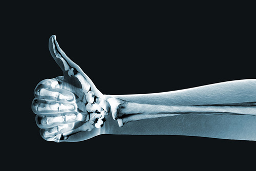
The images achieved via x-ray are done so by the brief transmission of electromagnetic waves (radiation) through a specific area of your body and onto photographic film. Bones, tumors, and other dense matter appear white (or light) on the x-ray – this is because those structures absorb radiation as opposed to allowing it (the radiation) to pass through to the film. It’s because of this that, once the x-ray film is developed, the exposure is one that reflects the structures within you.
Less dense areas of the body – such as soft tissues and/or breaks within a bone – allow the radiation to pass through, which is why these areas appear darker on the x-ray film. If a physician is trying to make certain organs stand out in the picture, you may be asked to drink a contrast, such as barium, which absorbs the radiation much like bone would.
X-rays are typically taken from several different angles to provide the physician with the most amount of views possible. If the x-ray is of a limb, a comparison x-ray may be taken of the unaffected/uninjured limb. X-ray sessions don’t tend to take long, and the images are usually ready quickly. It should be noted that, while the level of radiation exposure from a single x-ray isn’t harmful, if you are pregnant – or could be pregnant – your physician and x-ray technician will take additional precautions, just to be safe.
X-rays are a great initial diagnostic tool; however, they may not provide as much detail as newer, more powerful methods such as CT and MRI.
CT SCAN (COMPUTED TOMOGRAPHY)
A CT scan is a relatively modern imaging tool that combines X-rays with computer technology to produce more detailed, cross-sectional images of your body. A CT scan allows your medical team to see the size, shape, and position of the structures that are deep inside your body, such as organs, tissues, or tumors.
How Does a CT Scan Work?
When you have a CT, you’ll lay down on a table that slides into the center of a donut-shaped CT scanner. An x-ray tube slowly rotates around you, taking a multitude of images from all directions. Once the scan is complete, a computer then works to combine all the images into a clear, two-dimensional rendering that can be viewed on a screen.
This type of imaging can be used to view structures such as your brain and spinal cord, as well as organs within your chest, abdomen, and pelvis, or for providing more detailed images for small, bony structures. As with an X-ray, sometimes you may be given barium sulfate or a dye to make certain areas of your body show up better.
MRI (MAGNETIC RESONANCE IMAGING)
MRI’s are another modern imaging technique that are able to produce high quality, detailed cross-sectional images of your body. Unlike CT scans and x-rays, MRI’s do not use radiation. Instead, an MRI tool uses magnetic fields – as well as a sophisticated computer – to take high-resolution pictures of your bones and soft tissues. Because high-powered magnets are used, it’s extremely important to inform your doctor of any implants such as a pacemaker, metal clips, or other metal objects in your body before you undergo an MRI scan.
How Does an MRI Work?
When you have an MRI, you’ll lie on a table that slides into a tube-shaped MRI scanner. The MRI is then able to create a magnetic field around you, which pulses radio waves (which causes the affected tissue(s) to resonate) to the area of your body that needs to be imaged.
A computer then records the rate at which the various parts within your body – such as tendons, ligaments, nerves, etc. – give off these vibrations. These are then translated into a highly detailed, two-dimensional picture. You will not feel any pain while undergoing an MRI, but the machine may be noisy.
One of the potential limiting factors of an MRI was whether or not the patient could handle being inside the tube for an extended period of time – having an MRI isn’t a quick procedure (it typically takes between 60 and 90 minutes), which can pose serious problems for those who suffer from claustrophobia. However, the advent of “open” MRI machines has virtually eliminated this issue.
MRI’s a very helpful tool in diagnosing injury to cartilage, herniated disks, torn ligaments (like those in your knee), tears in the rotator cuff, hip and pelvic problems, as well as a multitude of other issues.
Obviously, there are a lot of options when it comes to diagnostic imaging options. Here at Prairie Orthopaedic, we have access to all the latest imaging options! If you’re struggling with an orthopaedic issue, please don’t hesitate to give us a call. Our physicians will discuss all your treatment options with you, including whether or not they feel that an X-ray, CT, or MRI will aide in your diagnosis.